NDT
“To create the 360˚ version of the
image the target is rotated along
with the multiple pulses of neutrons”
part of the image creation process. The boron plate
releases electrons when struck by the scattered neutrons
and the electrons are detected. “We amplify the electron
signal. We have a high frequency detector and the
image’s contrast varies based on the scanned object’s
microstructure. We have been doing this for a couple of
years,” Bilheux says.
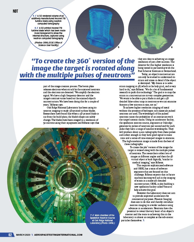
Oak Ridge National Laboratory has been using its
neutron imaging to study 3D-printed turbine blades.
Researchers have found that when a 3D printed blade is
cut from the build plate, the blade’s shape can subtly
change. The blades have been imaged to a resolution of
50 microns using their equipment and Bilheux says that
62 MARCH 2020 \\ AEROSPACETESTINGINTERNATIONAL.COM
they are close to achieving an image
resolution of just a few microns. The
detector for this higher resolution is
being tested in partnership with the
Paul Scherrer Institute in Switzerland.
Today, an object’s microstructure
can only be studied to understand its
strain and stress in detail if the object
is destroyed. “My dream is to solve
strain mapping in 3D, which is the holy grail, and is very
hard to do,” says Bilheux. “We do a lot of fundamental
research to push this technology.” The goal is to map the
strain in a microstructure in very complex geometries.
“We want to be able to put a blade in and get a 3D
detailed false-colour map in neutrons so we can examine
features a few microns in size, not 50.”
To achieve higher resolution imaging of metal
without the presence of hydrogen rich materials pulsed
neutrons are used. The wavelength of the pulsed
neutrons raises the probability of an interaction with
the target’s atomic nuclei. Using its accelerator facility,
the spallation neutron source, engineers at Oakridge
generate 60 pulses of neutrons per second and for each
pulse they have a range of neutron wavelengths. They
will produce about 2,000 radiographs from these pulses
and collect enough so they have good signal to noise
ratio and a series of time-stamped images to examine.
The high-resolution image is made from the best of
these radiographs.
To create the 360˚ version of the image the
target is rotated along with the multiple pulses
of neutrons. The researchers collect lots of
images at different angles and then the 3D
virtual object is built digitally, “similar to
medical imaging,” says Bilheux.
This requires sophisticated software
and ORNL has a team of software
engineers that are focused on this
challenge. Bilheux expects that in future
machine learning will aid in the imaging
of strain in a material’s detailed
microstructure. ORNL is also building a
new spallation facility called Venus to
help achieve this goal.
However the Laboratory does not aim
to provide improved accelerators for
commercial purposes. Phoenix Imaging
does aim to do that and thereby introduce
neutron imaging to a wider audience using
advances in accelerators. Neutrons have the
potential to reveal the very heart of an object’s
interior and the route to achieving this in the
laboratory is almost as complex as the sub-atomic
particles themselves. \\
7 // Main chamber of the
Spallation Neutron Source
at Oak Ridge National
Laboratory (Photo: ORNL)
5 // A 3D rendered volume of an
additively manufactured Inconel 718
turbine blade using neutron
computed tomography
6 // A 3D printed Inconel 718
turbine blade which has been made
more transparent to show the
internal structure, captured using
neutron computed tomography
(Photos: ORNL/DOE Office of
Science User Facility)
5 6
7
/AEROSPACETESTINGINTERNATIONAL.COM